
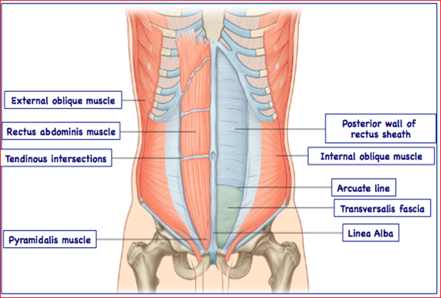
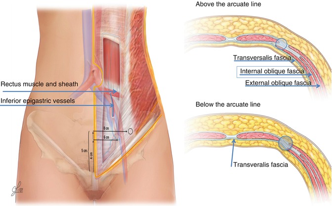
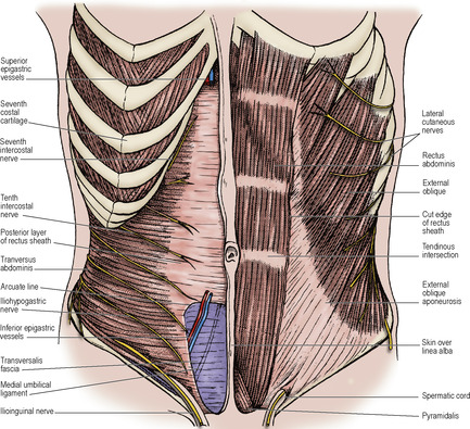
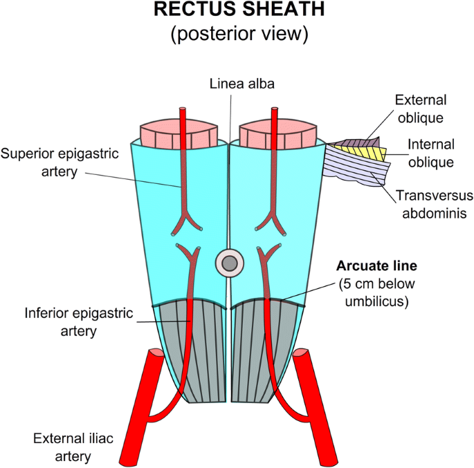
The arcuate line of rectus sheath the linea semicircularis the arcuate line or the semicircular line of Douglas is a horizontal line that demarcates the lower limit of the posterior layer of the rectus sheathIt is commonly known simply as the arcuate lineIt is also where the inferior epigastric vessels perforate the rectus abdominis. Thoracic paravertebral block TPVB is the technique of injecting local anesthetic alongside the thoracic vertebra close to where the spinal nerves emerge from the intervertebral foramen.
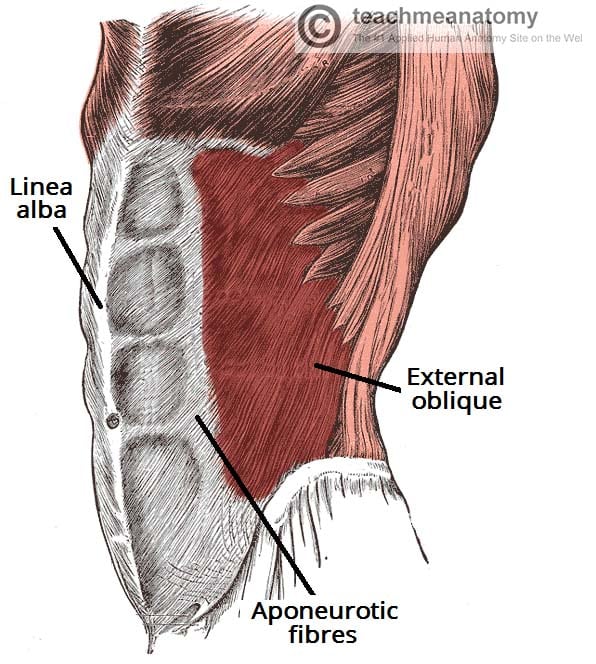
The Anterolateral Abdominal Wall Muscles Teachmeanatomy
1
016 Anterior Abdominal Wall Flashcards Memorang
The sacroiliac joints are essential for effective load transfer between the spine and the.
Arcuate line abdomen. This is called a Mullerian anomaly and can lead to many variants ranging from a uterine. This section features the relevant anatomy indications and technique descriptions to perform an ultrasound-guided TAP and QL plane blocks. Surface Anatomy Many of the organs in the abdominal cavity can be palpated through the abdominal wall or their position can be visualised by surface markings.
It is formed by the fusion of the aponeuroses of the muscles of the anterior abdominal wall. The adrenal glands sit immediately superior to the. This produces unilateral segmental somatic and sympathetic nerve blockade which is effective for anesthesia and in treating acute and chronic pain of unilateral origin from the chest and.
Popliteus muscle Musculus popliteus The popliteus muscle is a small muscle that forms the floor of the popliteal fossaIt belongs to the deep posterior leg muscles along with tibialis posterior flexor digitorum longus and flexor hallucis longus. The kidneys lie retroperitoneally behind the peritoneum in the abdomen either side of the vertebral column. Course It is typically a short vessel that passes underneath the median arcuate ligament often indented on its superior surface by this ligament and then courses anteriorly or slightly.
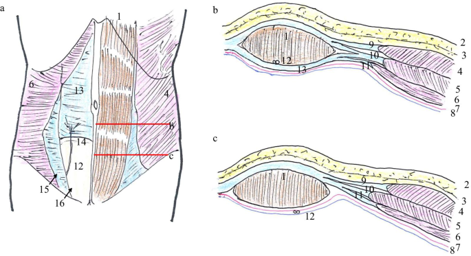
Ultrasound-guided transversus abdominis plane TAP and quadratus lumborum QL blocks have become a common analgesic method after surgery involving the abdominal wall. Sometimes the development in utero may be incomplete. The only exception is the posterior side of the lowest fourth of the rectus abdominis muscle below the arcuate line which is covered only by the transversalis fascia and parietal peritoneum.
Brief Reports and Innovations is a gold open access journal launched by Annals of Vascular Surgery. Each innominate is formed by the fusion of the three bones of the pelvis. On a coronal cut section its cavity has an inverted triangle shape.
Extraperitoneal fat and peritoneum. The popliteus muscle extends over the posterior aspect of the knee jointIt originates from the femur and the. The primary function of this artery is.
The new surgical journal seeks high-quality case reports small case series novel techniques and innovations in all aspects of vascular disease including arterial and venous pathology trauma arteriovenous. New Journal Launched. Immediately deep to the rectus sheath is the transversalis fascia below which lie the two deepest layers of the abdominal wall.
The aorta originates from the left ventricle of the heart. Arises anteriorly from abdominal aorta just below diaphragm at the T12 level behind the median arcuate ligament just as the aorta enters the abdomen in between right and left crura. Handy mnemonics to recall the contents of the spermatic cord are.
The Sacroiliac joint simply called the SI joint is the joint connection between the spine and the pelvis. Large diarthrodial joint made up of the sacrum and the two innominates of the pelvis. The uterus is a hollow pear-shaped organ that is responsible for a variety of functions such as gestation pregnancy menstruation and labor and delivery.
The demarcation point where the posterior layer of the rectus sheath ends is the arcuate line. They typically extend from T12 to L3 although the right kidney is often situated slightly lower due to the presence of the liverEach kidney is approximately three vertebrae in length. The ilium ischium and pubic bone.
In humans the linea alba runs from the xiphoid process to the pubic symphysis down the midline of the abdomenThe name means white line as it is composed mostly of collagen connective tissue which has a white appearance. The femoral artery is one of the major arteries in the human body. It ends in the abdomen where it branches into the two common iliac arteries.
Annals of Vascular Surgery. It continues sideways climbing over the sacroiliac joints along the iliopectineal lines in their initial portion linea innominata arcuate lines of the iliac bone also called innominate and continuing toward the pectineal line of the pubic bone crests and closing before the pubic symphysis passing through the superior-posterior edge of the iliopubic branches and the pubic. It separates the left and right rectus abdominis.
The descending aorta. Papers Dont Contribute To A Good Specialist Level 3 arteries 3 nerves 3 fascias 3 other things Mnemonics Papers Dont Contribute To A Good Specialist Level P. It extends from the iliac artery near the abdomen down to the legs.
The aorta has five separate segments.
Pubs Rsna Org
Basic Principles And Anatomy For The Laparoscopic Surgeon Abdominal Key
Arcuate Line

Arcuate Semi Circular Line When The Aponeuroses Of All Three Lateral Abdominal Muscles Pass Anterior To Abdominal Rectus Abdominis Muscle Abdominal Muscles
Single Incision Laparoscopic Repair For An Arcuate Line Hernia A Case Report Surgical Case Reports Full Text
Abdomen Basicmedical Key
Giant Rectus Sheath Hematoma The Ultrasound Journal Full Text

Figure Rectus Sheath Anatomy Superior And Statpearls Ncbi Bookshelf


